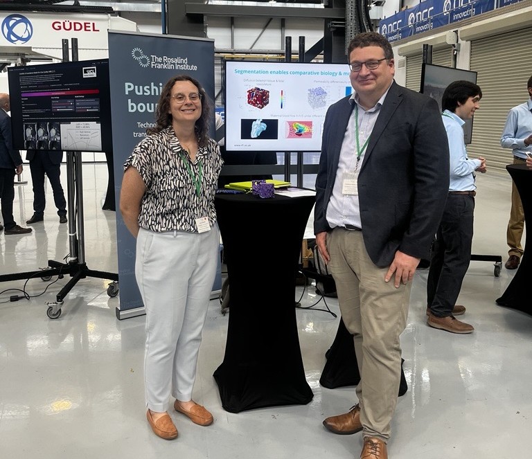
Harnessing the power of supercomputing to understand placental structure and its effect on pregnancy outcomes
The Rosalind Franklin Institute’s Advanced Research Computing team are using the UK’s latest supercomputer to help understand placenta imaging data. The time on the supercomputer will support multiple institute projects to help us understand the placenta and predict important conditions affecting pregnancy, including pre-eclampsia, fetal growth restriction or stillbirth.
The Franklin team were excited to have been granted early access to the UK’s newest supercomputer, Isambard-AI, which will significantly speed up data processing, enabling science that otherwise would not be possible.
“Access to Isambard-AI has changed our approach to image analysis as it opens up possibilities to scale the data differently,” said Dr Laura Shemilt, Head of Research Software Engineering at the Franklin.
The new £225m Isambard-AI facility, developed by the University of Bristol in close partnership with HPE and NVIDIA, is a key part of the UK Government’s AI Research Resource (AIRR), intended to boost the country’s capabilities in responsible and cutting-edge AI development.
The supercomputer, which is built and run by the Bristol Centre for Supercomputing (BriCS) was officially launched by Peter Kyle, the Secretary of State for Science, Innovation, and Technology.
The Franklin team were delighted to be at BriCS for the Isambard-AI launch. “It is a privilege to work with the Bristol Centre for Supercomputing on state-of-the-art computing for science. Big science creates big data sets, and to make meaning from them the UK needs these major centres for supercomputing” added Laura.

One of the projects the Franklin will be working on is an internationally collaborative project to reduce pregnancy complications, including stillbirths, funded by Wellcome Leap – In Utero.
The placenta is an organ which acts as an intermediary between the maternal and fetal circulation during pregnancy, changes to its structure can cause shifts in oxygen and nutrient transfer impacting the pregnancy outcome.
The Franklin provides expertise in segmentation; the process of image analysis where every pixel in an image is labelled and key structures are identified and quantified. The team are analysing 3D X-ray images of placental tissue collected at Diamond Light Source. When analysed by hand, this is an intensive and time-consuming process that severely limits researchers’ ability to look at a large enough number of samples to see meaningful differences in the biology.
“In these projects, we are using Isambard-AI to speed up our approaches to image analysis of X-ray and electron microscopy data so that we can learn which structural features are most important in the context of poor pregnancy outcomes. Then we’re using this information to find ways to identify these features in the clinic during pregnancy so that we can reduce the number of babies and mothers affected by pre-eclampsia, fetal growth restriction or stillbirth.
The imaging analysis pipelines that we are developing are not just useful in this context but could be adapted to work across imaging modalities and to tackle other pressing health challenges. Key to this work is the application of these approaches in areas where we can transfer what we learn from research into insights that can be used in the clinic to change how doctors treat patients,” said Dr Michele Darrow, Quantifying Biology across Scales Challenge lead.
The second project is a collaboration between the Franklin and University of Southampton, which aims to understand the evolutionary pressures that lead to structural variation in mammalian placentas.
The team at the Franklin have developed novel deep learning pipelines that enable semi-automated segmentation of serial block face scanning electron microscopy (SBF-SEM) datasets of human and animal placentas collected. By understanding placental structures, we can shed a light on the evolutionary reasons why humans are are the only mammals that suffer pregnancy-related conditions, such as pre-eclampsia or morning sickness. This work will also provide insights into the structure and function of placentas across various animal species potentially providing insights that could lead to improved animal husbandry or conservation efforts by improving pregnancy outcomes for domestic (e.g., cattle, sheep) or exotic (e.g., rhino, giraffe) animals.
